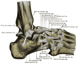
Ankle inversion sprains are a very common injury seen in the primary care setting. The vast majority of ankle sprains are treated nonoperatively with appropriate conservative treatment. After a brief period (1-2 weeks) of rest, ice, compression, and elevation, most patients are able to progressively return to their activities. A small minority of patients continue to have persistent instability and require operative intervention to repair their lateral ligaments, most commonly referred to as a Brostrom procedure.
 Ankle Pain Physical Examination
In the patient with chronic instability (not the acutely presenting patient), inspection should focus on persistent swelling or ankle effusion. Additional items include: Range of motion of both the ankle and the subtalar joint, tenderness to palpation about the ankle and peroneal tendons, as well as eversion and inversion strength. Laxity testing of the ankle consists of the anterior drawer and talar tilt tests. The anterior drawer checks for laxity in the anterior talofibular ligament and is done by placing the ankle in 20 degrees of flexion, grasping the heel, pulling forward, and noting the amount of translation. The talar tilt test checks for laxity in the calcaneofibular ligament and is done in neutral flexion, grasping the heel and inverting the hindfoot. Obviously, both tests are done on the opposite sides for comparison.
Ankle Pain Imaging
Most patients with chronic ankle instability should undergo a standard three view ankle x-ray series. The x-rays should be scrutinized for osteochondral lesions of the talus or tibial plafond. Osteochondral lesions, persistent pain in the peroneal tendon area, and chronic ankle effusions are indications for a noncontrast MRI of the ankle. This can be requested at the same time as an orthopedic consultation. In a straight-forward case of persistent ankle instability, an MRI is not required.
